Anatomy Label Major Arteries And Veins / Vein Blood Vessel Britannica : Human anatomy for muscle, reproductive, and skeleton.. Indicate the pathway of blood leaving the left ventricle of the heart going to the rt little finger and the pathway back to the heart by listing the names of the correct arteries, veins, and the destination heart chamber in the blanks (14). Arteries typically have a thicker tunica media than veins, containing more smooth muscle cells and elastic tissue. The external carotid artery supplies the areas of the head and neck external to the cranium. Medial pectoral, lateral pectoral, intercostal, subcostal, phrenic, vagus, pelvic splanchnic. This artery stems from the axillary artery.
Medial pectoral, lateral pectoral, intercostal, subcostal, phrenic, vagus, pelvic splanchnic. Learn anatomy faster and remember everything you learn. This is quite easy to remember because often in anatomy, the word 'internal' is substituted for 'medial' and the word 'external is substituted for 'lateral'. There are about half a dozen arteries to learn. Anatomy and physiology questions and answers.
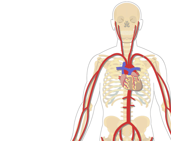
Art Labeling Quiz from wps.pearsoned.com The external carotid artery supplies the areas of the head and neck external to the cranium. Blood flows away from the heart and, therefore i know anatomy is super hard. Describe the waveforms and pressures that are seen in each anatomical location during insertion of a pulmonary artery catheter. Thoracic aorta, abdominal aorta, iliac arteries veins: Explore the anatomy of the human cardiovascular system (also known as the circulatory system) with our detailed diagrams and information. General anatomy and musculoskeletal system. Anatomy visible in the medical illustration includes: Arteries typically have a thicker tunica media than veins, containing more smooth muscle cells and elastic tissue.
Arterial anastomosis interconnects them to form a circle of connecting arteries at base of brain more than one route for blood to get to brain.
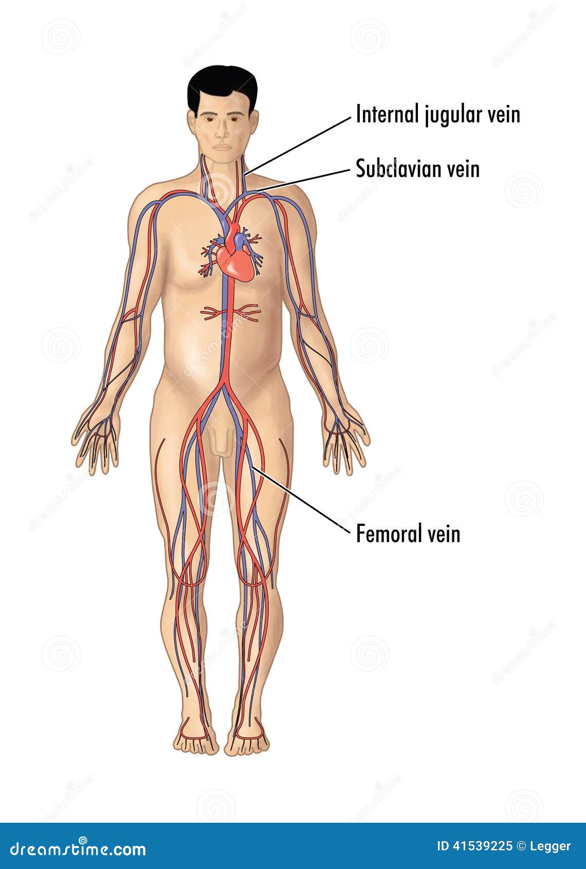
Arteries carry oxygenated and nutrient rich blood to the bodys tissues from the heart. Arteries carry oxygenated blood (with the exception of the pulmonary artery and umbilical artery). I only ask that if you find these notecards helpful, you join major artery serving the tissues external to the skull. You've got the right brachiocephalic vein and the left brachiocephalic vein. Match the arteries in column a with the regions supplied in column b. Veins are blue blood vessels that carry blood towards the heart. Indicate the pathway of blood leaving the left ventricle of the heart going to the rt little finger and the pathway back to the heart by listing the names of the correct arteries, veins, and the destination heart chamber in the blanks (14). Arteries and veins of the human body. Anatomy visible in the medical illustration includes: Goes though both pec major obturator nerve artery vein. Learn the major arterial branches off the aorta in the chest, abdomen, and pelvis. Anatomy and physiology questions and answers. Roots, trunks, divisions, cords, branches.
Diffen › science › biology › anatomy. This artery stems from the axillary artery. Thoracic aorta, abdominal aorta, iliac arteries veins: Learn anatomy faster and remember everything you learn. This illustration was published in.
Major Systemic Arteries from www.getbodysmart.com Describe the waveforms and pressures that are seen in each anatomical location during insertion of a pulmonary artery catheter. There are about half a dozen arteries to learn. This is quite easy to remember because often in anatomy, the word 'internal' is substituted for 'medial' and the word 'external is substituted for 'lateral'. Illustration depicting main leg arteries (anterior view). Together, veins, arteries and nerves define neurovasculature. Anatomy of the arterial wall : Electrical properties of the heart. Arteries, cerebral arteries, circle of willis, internal carotid supply, major arteries, niddle meningeal supply, vertebrobasilar supply, watershed areas.
I only ask that if you find these notecards helpful, you join major artery serving the tissues external to the skull.
Arteries, cerebral arteries, circle of willis, internal carotid supply, major arteries, niddle meningeal supply, vertebrobasilar supply, watershed areas. Simple labelled illustration depicting the general pathways for the major arteries of the head and neck. There are three major types of blood vessels: This illustration was published in. Major arteries, pulse points, and veins. Describe the waveforms and pressures that are seen in each anatomical location during insertion of a pulmonary artery catheter. Veins need valves to create pressure to pump the blood to the heart. Anatomy of excitatory and conductive elements: Blood vessels 1, arteries and veins. Learn anatomy faster and remember everything you learn. Last updated on sat, 03 apr 2021 | human anatomy. Anatomy and physiology questions and answers. Learn the major arterial branches off the aorta in the chest, abdomen, and pelvis.
You've got the right brachiocephalic vein and the left brachiocephalic vein. General anatomy and musculoskeletal system. Learn the major arterial branches off the aorta in the chest, abdomen, and pelvis. Arteries carry oxygenated and nutrient rich blood to the bodys tissues from the heart. This illustration was published in.
Major Blood Vessels For Central Line Insertion Stock Illustration Illustration Of Science Medical 41539225 from thumbs.dreamstime.com Arteries carry oxygenated blood (with the exception of the pulmonary artery and umbilical artery). Lateral pectoral nerves goes through pectoralis major while medial p.n. Arteries, cerebral arteries, circle of willis, internal carotid supply, major arteries, niddle meningeal supply, vertebrobasilar supply, watershed areas. Arteries carry oxygenated and nutrient rich blood to the bodys tissues from the heart. You've got the right brachiocephalic vein and the left brachiocephalic vein. Electrical properties of the heart. There are three major types of blood vessels: Learn the major arterial branches off the aorta in the chest, abdomen, and pelvis.
Major branches (medial portions of frontal lobes, superior medial part of parietal.
15.1 abdominal aorta and major branches anterior view. Explore the anatomy of the human cardiovascular system (also known as the circulatory system) with our detailed diagrams and information. This artery stems from the axillary artery. Head, neck, arteries, external carotid, internal carotid, common carotid, temporal, occipital, posterior auricular, carotid sinus, vertebral. Describe the waveforms and pressures that are seen in each anatomical location during insertion of a pulmonary artery catheter. Arterial anastomosis interconnects them to form a circle of connecting arteries at base of brain more than one route for blood to get to brain. Arteries carry oxygenated blood (with the exception of the pulmonary artery and umbilical artery). Together, veins, arteries and nerves define neurovasculature. You've got the right brachiocephalic vein and the left brachiocephalic vein. Related posts of anatomy veins arteries diagram. Arterial wall layers including the tunica intima and the tunica media. Illustration depicting main leg arteries (anterior view). Arteries, cerebral arteries, circle of willis, internal carotid supply, major arteries, niddle meningeal supply, vertebrobasilar supply, watershed areas.


0 Komentar